In this section of the PALS course, we're going to cover the cardiac conduction system and all of its components, and we'll start with a deep dive into the biomechanical and electromechanical actions of the heart.
In this lesson, we'll take a closer look at the heart and how it functions as a circulatory muscle, including the mechanisms that allow it to function.
The myocardium is a muscle unlike any other muscle that we have in our bodies. What makes it so unique is its ability to generate its own electrical impulses, known as automaticity.
Pro Tip #1: Automaticity is the body's ability to do things without occupying the mind and with low-level details required, allowing it to become an automatic response pattern or habit.
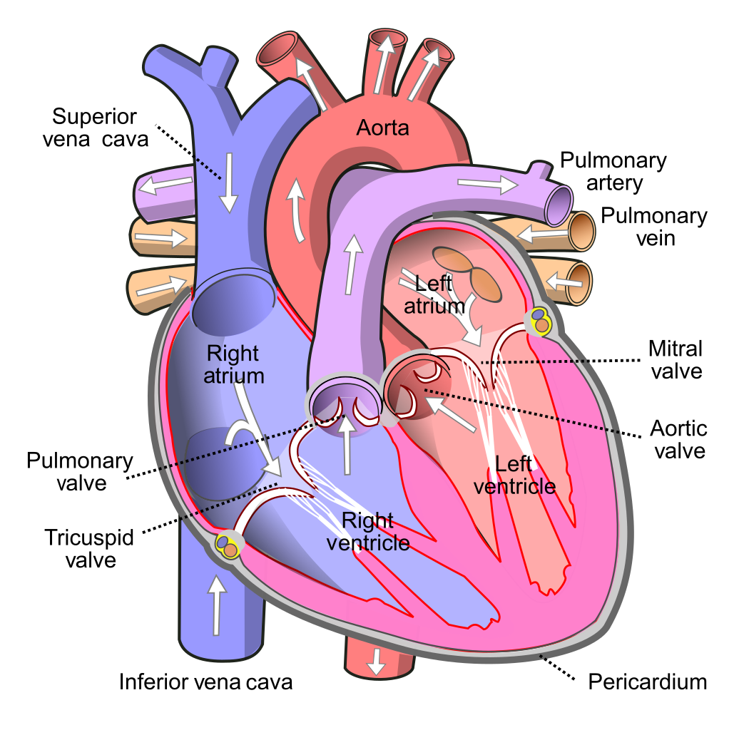 One particularly special part of the heart muscle is located in the superior aspect of the right atrium, called the sinoatrial node, or SA node for short. It works like an internal/biological pacemaker.
This SA node, when the heart is functioning as it was designed to function, generates an electrical impulse that travels through the myocardium in a very organized and deliberate way. The SA node generates electrical impulses at a rate between 60 and 100 times per minute.
If we were to follow the pathway of that electrical impulse from the SA node to the place where it terminates, that place would be at the end of the Purkinje fibers.
Pro Tip #2: The Purkinje fibers are specialized conducting fibers composed of electrically excitable cells that are larger than cardiomyocytes with fewer myofibrils and many mitochondria and which cells conduct cardiac action potentials more quickly and efficiently than any other cells in the heart.
After the SA node initiates that electrical impulse, it then travels via pathways, known as internodal pathways, throughout the right and left atria. It then depolarizes the myocardia cells which causes the heart muscle in the atrium to contract.
From the atria, that electrical impulse travels along the pathway to the atrial ventricular node, or the AV node, where it's strategically delayed before moving through the bundle of His, or AV bundle, and ultimately to the Purkinje fibers.
The Purkinje fibers travel down through and around the ventricles, thereby completing the electromechanical cycle of one complete heartbeat.
The delay in the AV node, which is located in the left lower wall of the right atrium, is a very necessary process. This delay allows the ventricles to beat independently of one another, which allows them to operate as a double pump action.
If for whatever reason, the SA node doesn't operate properly as the primary impulse generator, or our biological pacemaker, the AV node can then begin sending its own electrical impulse instead; providing the heart with a failsafe mechanism or backup electrical generator.
While the AV node can generate its own electrical impulses, it does so at a much slower rate, which ranges between 40 and 60 impulses per minute.
When the AV node is called upon to generate this electrical impulse, it travels from the AV node through the bundle of His and eventually reaches the Purkinje fibers, which wrap around the ventricles we mentioned earlier, and once again completing the electromechanical cycle of one complete heartbeat.
This ventricle contraction then circulates the majority of oxygenated blood throughout the rest of the body.
Pro Tip #3: The bundle of His is the bundle of cardiac muscle fibers that conducts the electrical impulses that regulate the heartbeat, from the AV node in the right atrium to the septum between the ventricles, and then to the left and right ventricles.
Upon reaching the bundle of His, that electrical impulse then travels down the length of the intraventricular septum, which leads to the left and right bundle branches. The left bundle branch has two fascicles (or bundle of fibers) due to its size, since the left ventricle is larger than the right ventricle, which has only one fascicle.
These bundle branches ultimately terminate, or lead into, the Purkinje fibers, which then depolarize the ventricular cells and cause the ventricular muscles to contract.
In situations where both the SA and AV nodes aren't able to generate electrical impulses properly, the Purkinje fibers located within the ventricles then become the primary pacemaker source. The problem with this scenario is that the Purkinje fibers only generate electrical impulses in the range of around 15 to 40 beats per minute.
This rate is usually too slow to produce adequate systolic blood pressure or oxygenate the cells in the body.