Atrial flutter (AFL) is a common abnormal heart rhythm that starts in the atrial chambers of the heart. When it first occurs, it is usually associated with a fast heart rate.
In this lesson, we'll look at why/how atrial flutter occurs, and then look at a typical ECG readout for an adult patient in atrial flutter and provide a cardiac interpretation at the end.
On an ECG, atrial flutter typically includes sawtooth-like F-waves, which are either the result of an ectopic atrial pacemaker or because of rapid reentry pathways somewhere within the atria, but outside of the SA node.
The origin of this ectopic pacemaker is usually somewhere in the lower atrium and closer to the AV node, thereby resulting in that distinct sawtooth wave pattern.
Pro Tip #1: Due to this erratic electrical activity, the normal function of the SA node is usually suppressed and noneffective. Which is why, instead of a P-wave, atrial flutter will produce flutter, or F-waves. And as a result of the depolarization of the atria in an abnormal manner, the classic F-waves of atrial flutter resemble a sawtooth, hence the name.
Now let's take a look at an ECG for an adult patient in atrial flutter.
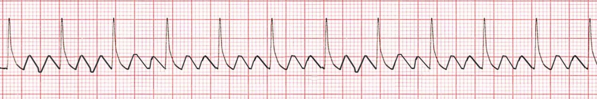
*Atrial Flutter ECG for Adult Patient
1. The Heart Rhythm
The first thing you'll want to look at is the heart rhythm. Does the heart rhythm look regular? Or does it look irregular? In the ECG above, the rhythm is variable and dependent on the ratio of F-waves to the QRS complexes.
2. The Heart Rate
Next, you'll want to look at the heart rate of the patient. What is the patient's heart rate? Is it normal? Or is it too slow or too fast? In this case, it's variable due to its irregularity.
3. P-Wave
After looking at the heart rate, check to see if the patient's P-waves look normal by asking yourself the following few questions.
- Are the patient's P-waves present, and do they resemble normal P-waves or just those sawtooth type of F-waves?
Since the answer is, they resemble sawtooth style F-waves, all of the other P-wave questions you normally ask yourself do not apply, once you notice the F-wave flutter. There are no real SA node P-waves present.
4. PR Interval
Next, look at the PR interval on the patient's ECG readout and ask yourself the following questions:
- Is the PR interval normal, meaning between .12 and .20 seconds or is it contained within one large square on the readout? The answer is no, because it's variable and there are no P-waves.
- Is the PR interval constant? Again, this is non-applicable because of the above answer.
5. QRS Complex
The last thing you should look at to determine if the sinus rhythm is normal or not is the QRS complex and ask yourself these questions while you do:
- Is the QRS interval less than .12 seconds? Yes, it is within the normal range.
- Is the QRS complex wide or narrow? In this case, it's narrow.
- Are the QRS complexes similar in appearance or are there noticeable differences? In this case, we can see that each looks similar.
So, what is your cardiac interpretation? Based on these questions and on the findings from the ECG readout above, it would appear that this patient is in atrial flutter.
- We have a variable rhythm that is dependent on the ratio of F-waves to the QRS complexes.
- We have a variable heart rate due to its irregularity.
- The P-waves are not normal and resemble sawtooth style F-waves.
- The PR interval is variable and there are no normal P-waves.
- The QRS is less than .12 seconds and thus normal.
From the ECG alone, it would indicate that the patient is in atrial flutter
Pro Tip #2: Structural heart disease is the usual cause of atrial flutter. In the same way that atrial fibrillation complicates adequate ventricular preload filling, atrial flutter complicates circulation and especially when it is accompanied by a syndrome called rapid ventricular rate or response.
What is rapid ventricular rate or response? In some cases of AFib, the fibrillation of the atria causes the ventricles, or lower chambers of the heart, to beat too fast. When this happens, it's called a rapid ventricular rate or response, or RVR for short.
Pro Tip #3: The faster the ventricular response, the more likely it is that the patient's circulation will be compromised. When the ventricles beat too rapidly, they aren't able to fill completely with blood from the atria. As a result, they can't efficiently pump blood out to meet the needs of the body. This can ultimately lead to heart failure.