Supraventricular tachycardia, or SVT, refers to a tachycardia originating above the bundle of His, typically involving a reentrant circuit within or near the AV node. Unlike normal sinus rhythm, where the SA node drives a controlled, regular rate, SVT involves an abnormal electrical circuit that bypasses or overrides that normal pacemaker mechanism, resulting in a rapid heart rate.
In this lesson, we'll take a deeper dive into supraventricular tachycardia for the adult patient, including looking more closely at an example of what it looks like on an ECG and see what findings and measurements lead us to our conclusion.
Understanding Tachycardia
Tachycardia simply describes an abnormally fast heart rate. And there are a several different types of tachycardias – narrow complex or wide complex and regular or irregular.
To be more specific, if the QRS interval is less than 0.12 seconds in length, that signifies a narrow complex tachycardia, or SVT. If the QRS interval is greater than 0.12 seconds, that signifies a wide complex tachycardia, and we'll get more into wide complex tachycardias in a subsequent lesson.
However, just know that this is significant because it's measurable and can tell us if the cause is atrial-based or ventricular-based.
If the patient's heart rate is too fast for their condition or the condition of their heart, the result is usually a decrease in cardiac output, poor perfusion of oxygenated blood, and a decrease in blood pressure.
Supraventricular Tachycardia (SVT)
With SVT, that stimulus comes from a rogue myocardial cell that stimulates an erratic atrial contraction, or a series of erratic atrial contractions, like those found in patient's with atrial fibrillation and atrial flutter.
Remember, Atrial fibrillation (also called AFib) is a quivering or irregular heartbeat (arrhythmia) that can lead to blood clots, stroke, heart failure, and other heart-related complications.
And as you know, Atrial flutter (AFL) is a common abnormal heart rhythm that starts in the atrial chambers of the heart. When it first occurs, it is usually associated with a fast heart rate.
Pro Tip #1: While these appear to be the same, the difference is in the beat. Atrial flutter and atrial fibrillation are both abnormal heart rhythms. However, in atrial fibrillation, the atria beat irregularly, while in atrial flutter, the atria beat regularly, but faster than usual and more often than the ventricles, so you may have four atrial beats to every one ventricular beat.
The important thing to note with SVT is that it can persist until there is medical intervention, or it can be intermittent and self-limiting and can come and go without warning.
By looking at an ECG readout alone, SVT can be difficult to differentiate from sinus tachycardia, AFib, or AFL. However, there are things that you can look at to help you determine which rhythm is being displayed.
Now let's take a look at an ECG for a patient with supraventricular, or narrow complex, tachycardia.
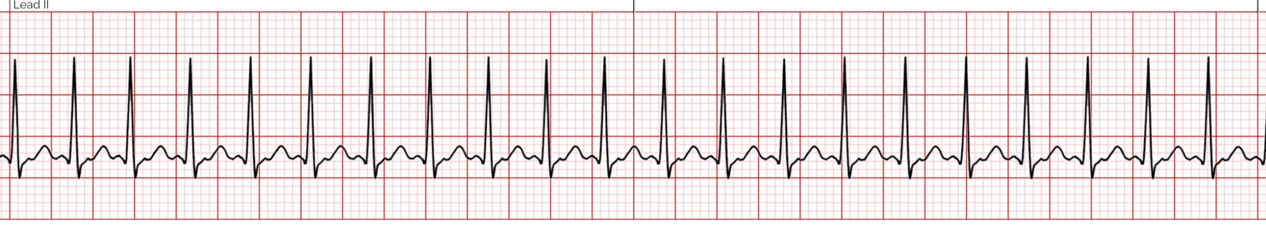 *Narrow Complex Supraventricular Tachycardia ECG for Adult Patient
1. The Heart Rhythm
The first thing you'll want to look at is the heart rhythm. Does the heart rhythm look regular? Or does it look irregular? In the ECG above, the rhythm is regular.
2. The Heart Rate
Next, you'll want to look at the heart rate of the patient. What is the patient's heart rate? Is it normal? Or is it too slow or too fast? In this case, it's too fast and greater than 100 beats per minute.
3. P-Wave
After looking at the heart rate, check to see if the patient's P-waves look normal by asking yourself the following few questions.
- Are the patient's P-waves present? Yes, the P-waves are present and upright.
4. PR Interval
Next, look at the PR interval on the patient's ECG readout and ask yourself the following questions:
- Is the PR interval normal for an adult patient, meaning between 0.12 and 0.20 seconds, or is it contained within one large square on the readout? Yes, it is.
- Is the PR interval constant? Yes.
5. QRS Complex
The last thing you should look at to determine if the sinus rhythm is normal or not is the QRS complex and ask yourself these questions while you do:
- Is the QRS interval less than 0.12 seconds? Yes, the QRS interval is between 0.06 and 0.11 seconds.
- Is the QRS complex wide or narrow? In this case, it's narrow.
Pro Tip #2: It's unusual for SVT to present with a wide complex QRS.
- Are the QRS complexes similar in appearance or are there noticeable differences? In this case, we can see that each looks similar.
So, what is your cardiac interpretation? Based on these questions and on the findings from the ECG readout above, it would appear that this patient is in supraventricular tachycardia.
- We have a regular rhythm.
- We have a faster than normal heart rate at greater than 100 beats per minute.
- The P-waves are present and upright.
- The PR interval is between 0.12 and 0.20 seconds.
- The QRS is between 0.06 and 0.11.
- The P:QRS ratio is 1:1.
From the ECG alone, it would indicate that the patient is in SVT. However, patient signs and symptoms must be taken into account to properly identify the rhythm correctly and to determine whether or not treatment is necessary.
The leading causes of most tachycardias are:
- Heart disease
- Electrolyte imbalance
- Medications
- Hypoxemia
- Other causes of hemodynamic instability
Regardless of the cause, if the patient is unstable, rapid treatment must be given immediately to correct the cause of the tachycardia.
Pro Tip #3: Keep in mind, a narrow complex tachycardia is less likely to cause hemodynamic instability and, in some cases, can be a normal response to the body requiring better circulation due to fear, exercise, or due to moderate bleeding resulting in blood volume issues.