In this lesson, we're going to look at the four types of atrioventricular blocks, usually called AV heart blocks or AV blocks for short. The four types are:
- 1st degree heart block
- 2nd degree heart block
- 2nd degree type 2 heart block
- 3rd degree heart block
We'll include an example ECG for each, so you can see the differences, while also reading about those differences.
1st Degree AV Heart Block
First-degree heart blocks are usually caused by a delayed, inconsistent, and sometimes absent electrical conduction pathway traveling through the AV node and can exhibit the following signs on an ECG readout.
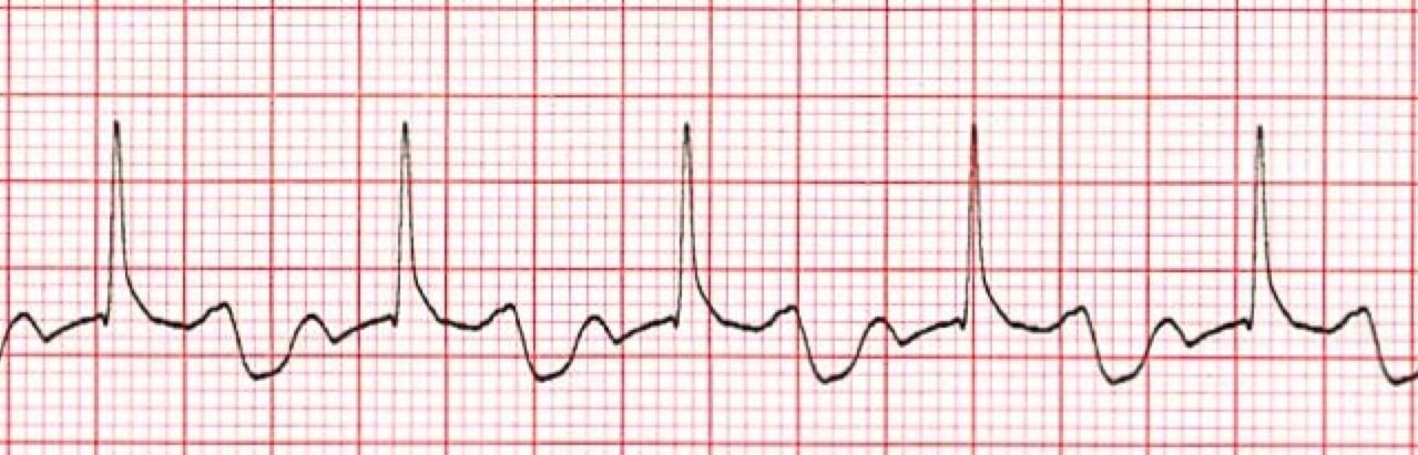 *1st Degree AV Heart Block ECG for Patient
| 1. | Rhythm | regular |
| 2. | Rate | normal or slow |
| 3. | P-waves | present and upright |
| 4. | PR interval | prolonged, beyond .20 seconds |
| 5. | QRS complex | between .06 and .11 seconds |
| 6. | P:QRS ratio | 1:1 |
There is usually little to no clinical significance with this type of heart block.
2nd Degree AV Heart Block (Mobitz Type 1)
Second-degree heart blocks, also known as Mobitz type 1 AV blocks, is commonly caused by:
- Heart disease affecting the AV node
- Vagal stimulation that's often associated with difficult bowel movements
- Coughing fits
- Certain medications
An ECG for a patient with Mobitz type 1 will exhibit the following signs.
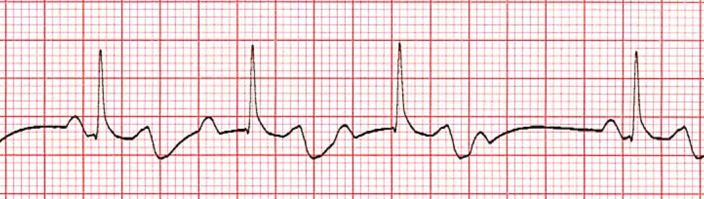
*2nd Degree (Mobitz type 1) AV Heart Block ECG for Patient
| 1. | Rhythm | regularly irregular |
| 2. | Rate | normal or slow |
| 3. | P-waves | present and upright |
| 4. | PR interval | progressively widening |
| 5. | QRS complex | between .06 and .11 seconds |
| 6. | P:QRS ratio | 1:1, until the P-wave is blocked |
Pro Tip #1: The QRS complex will become progressively delayed at the AV node until it completely disappears. When this happens, the ECG will only show a P-wave but no QRS following it.
2nd Degree AV Heart Block (Mobitz Type 2)
The third type of heart block is regularly known as a Mobitz type 2 block. It usually occurs when the heart block is below the AV node. A Mobitz type 2 block is usually caused by more advanced heart disease and can also originate from damage below the bundle of His.
Because of this, Mobitz type 2 can deteriorate more quickly into a symptomatic dysrhythmia and could eventually become a 3rd-degree heart block.
An ECG for a patient with Mobitz type 2 will appear to have intermittent blocks where some P-waves do not have a QRS complex following, and there's typically no elongation of the PR interval.
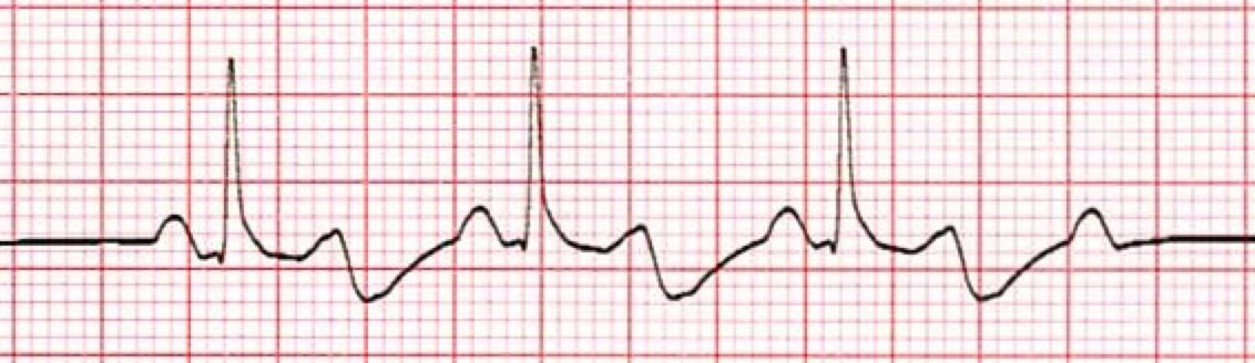
*2nd Degree (Mobitz type 2) AV Heart Block ECG for Patient
| 1. | Rhythm | variable, depending on the P:QRS ratio |
| 2. | Rate | variable, but usually slow |
| 3. | P-waves | present and upright |
| 4. | PR interval | between .12 and .20 seconds |
| 5. | QRS complex | between .06 and .11 seconds |
| 6. | P:QRS ratio | variable – 2:1, 3:1, 4:1 and greater |
3rd Degree AV Heart Block
The fourth and last type of heart block is called a 3rd degree complete AV heart block and is the most serious of the four.
A 3rd-degree heart block occurs when the electrical conduction is completely blocked between the atria and the ventricles. The exact location of the block can vary, however it's usually around the AV node or lower but will disassociate the SA pacemaker from the AV or bundle of His pacemakers.
Pro Tip #2: When this happens, a 3rd degree AV heart block will create an ECG readout that shows regular P-waves, regular QRS waves, but they'll be at different rates that are completely disassociated altogether.
An ECG for a patient with a 3rd-degree heart block will exhibit the following signs.
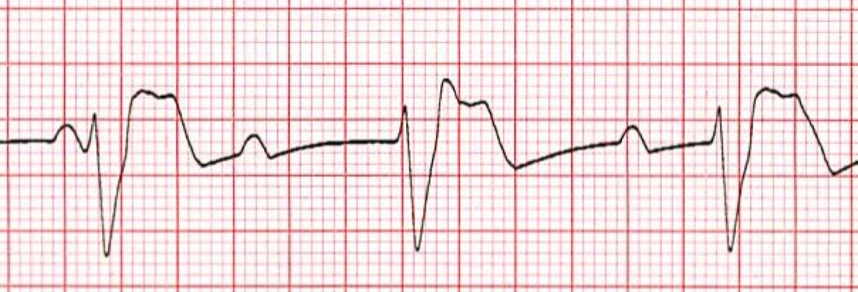
*3rd Degree AV Heart Block ECG for Patient
| 1. | Rhythm | regular |
| 2. | Rate | bradycardic and between 20 and 40 beats per minute |
| 3. | P-waves | present and upright |
| 4. | PR interval | variable with no set pattern |
| 5. | QRS complex | greater than .11 seconds |
| 6. | P:QRS ratio | variable |
The clinical significance of this type of dysrhythmia is serious. The patient will usually be symptomatic and unstable due to their very slow bradycardic heart rhythm and rate.
Pro Tip #3: This type of heart block is preventing any pace that originates from the SA node. Therefore, the ventricular pacemaker will stimulate a pulse rate closer to 20 to 40 beats per minute, which is usually not enough to maintain a stable blood pressure. This is why the ECG readout will usually display wide QRS complexes.
Studies have shown that 3rd degree AV heart blocks may be transient or permanent, depending on underlying causes.