


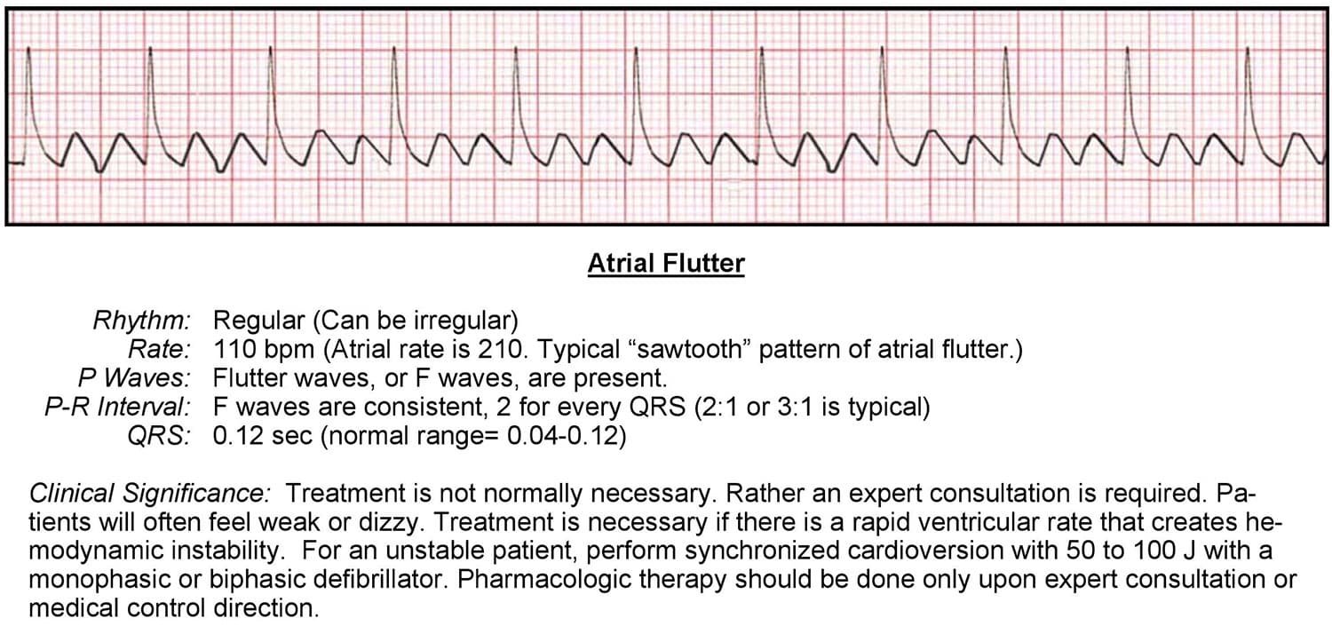
On an ECG, atrial flutter resembles F waves with a sawtooth pattern. This is the result of an ectopic atrial pacemaker or because of a rapid reentry pathway somewhere within the atria but outside of the SA node area.
This ectopic pacemaker usually originates somewhere in the lower atrium and closer to the AV node, making a distinct wave pattern. Due to this erratic electrical activity, the normal function of the SA node is usually suppressed and non-effective. So, instead of P waves, atrial flutter produces flutter (F) waves. Because of the abnormal depolarization of the atria, the flutter waves resemble a sawtooth pattern.

To interpret an ECG, ask the following questions:
Rhythm
- Is the rhythm regular or irregular?
- Variable, depending on the ratio of F waves to QRS complexes
Rate
- What is the rate?
- The rate is variable
- Is the rate normal, fast or slow?
- The rate is variable because of its irregularity
P Wave
- Are they present?
- Anything closely resembling a P wave is actually a sawtooth-type F wave
- Do they occur regularly?
- No, because there are no real SA node P waves
- Is there one P wave for each QRS complex?
- No, there is no P wave for each QRS complex
- Are the P waves smooth, rounded, and upright?
- No, they are Sawtooth in shape
- Do all P waves have similar shapes?
- No
PR Interval
- Does the PR interval fall within the normal 0.12-0.20 seconds?
- No, it’s variable
- Is the PR interval constant?
- No
QRS
- Is the QRS interval less than 0.12 seconds?
- Yes, the QRS is within normal range
- Is the QRS wide or narrow?
- In this case, the QRS is narrow
- Are the QRS complexes similar in appearance?
- Each one looks similar
Cardiac Interpretation
Atrial flutter is usually caused by structural heart disease. Similarly to how atrial fibrillation complicates ventricular preload filling, atrial flutter complicates circulation. This is especially true when atrial flutter is accompanied by a syndrome called rapid ventricular response. The faster the ventricular response, the more the circulation will be compromised.