Note: Your progress in watching these videos WILL NOT be tracked. These training videos are the same videos you will experience when you take the full ProACLS program. You may begin the training for free at any time to start officially tracking your progress toward your certificate of completion.
When talking about treating a patient for something that we consider abnormal, it's always helpful to define and understand what normal looks like, in this case, for a normal sinus rhythm.
In this lesson, we'll look more closely at an example of a normal sinus rhythm on an ECG (aka EKG) for an adult patient and see what findings and measurements are considered normal, and what to be on the lookout for that would be considered abnormal. And at the end of the lesson, we'll provide a Word about acute coronary syndrome.
 *Normal Sinus Rhythm ECG/EKG for Adult Patient
1. The Heart Rhythm
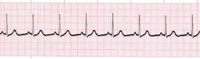
The first thing you'll want to look at is the heart rhythm. Does the heart rhythm look regular? Or does it look irregular? In the above graphic, it's regular.
2. The Heart Rate
Next, you'll want to look at the heart rate of the patient. What is the patient's heart rate? Is it normal? Or is it too slow or too fast?
Remember, to determine the patient's heart rate you'll want to observe the following areas on the ECG paper printout and perform the following calculations.
The horizontal axis of ECG paper grids is where time is measured. Each small square is 1mm in length and represents .04 seconds. Each larger square is 5mm in length and represents .2 seconds. Therefore a 6 second interval would be 30 large squares.
To determine the heart rate, count the number of QRS complexes over this 6 second interval and multiply by 10.
In the ECG above, the rate is 80 beats per minute, and this is normal. For an adult patient, the normal heart rate range is 60 to 100 beats per minute.
3. P-Wave
After looking at the heart rate, check to see if the patient's P-waves look normal by asking yourself the following few questions.
- Are the patient's P-waves present?
- Do they occur regularly?
- Is there one P-wave for each QRS complex?
- Are the P-waves smooth, rounded, and upright?
- Do all the P-waves have a similar shape?
The answer to each of those questions is, yes, meaning the P-waves are normal.
4. PR Interval
Next, look at the PR interval on the patient's ECG readout and ask yourself the following questions:
- Is the PR interval normal for an adult patient, meaning between .12 and .20 seconds, or is it contained within one large square on the readout?
- Is the PR interval constant?
The answer to both questions is, yes.
5. QRS Complex
The last thing you should look at to determine if the sinus rhythm is normal or not is the QRS complex and ask yourself these questions while you do:
- Is the QRS interval less than .12 seconds?
Pro Tip: As long as the QRS fits within two small squares on the ECG printout and is not greater than three small squares, it's within the normal range.
- Is the QRS complex wide or narrow? If it's narrow, such as on the ECG printout above, then that's considered normal.
- Are the QRS complexes similar in appearance or are there noticeable differences? For the above ECG readout, the answer is, they're similar in appearance and thus normal.
So, what is your cardiac interpretation? (This is something we'll be asking ourselves each time we look at a new ECG rhythm.) Based on these questions and on the findings from the ECG readout above, it's safe to say that the patient has a normal sinus rhythm.
- We have a regular rhythm.
- We have a normal heart rate.
- The P-waves look normal, with each being followed by a QRS complex.
- The PR interval is between .12 and .20 seconds.
- The QRS is less than .12 seconds.
Unless the patient has no pulse or other serious signs or symptoms, it's safe to assume that there is nothing of significance, in a negative sense, from this patient's cardiac rhythm.
A Word About Acute Coronary Syndrome
As an ACLS provider, you should have the basic knowledge to assess and stabilize patients with acute coronary syndrome (ACS). In these cases, you will use the ACS algorithm as your guide to clinical strategy.
The initial 12-lead ECG is used in all ACS cases to classify patients into one of three ECG categories. Each of these categories has different strategies of care and management needs.
The three ECG categories are ST-segment elevation suggesting ongoing acute injury, ST-segment depression suggesting ischemia, and nondiagnostic or normal ECG. All three are outlined in the ACS Algorithm.
Key components of these cases are:
- Identification, assessment, and triage of acute ischemic chest discomfort
- Initial treatment of possible ACS
- Emphasis on early reperfusion of the patient with ACS/STEMI (ST-Elevation Myocardial Infarction)
Rhythms for ACS
Sudden cardiac death and hypotensive bradyarrhythmias may occur with acute ischemia. You should learn to anticipate these rhythms and be prepared for immediate attempts at defibrillation and administration of medication or electrical therapy for symptomatic bradyarrhythmias.


